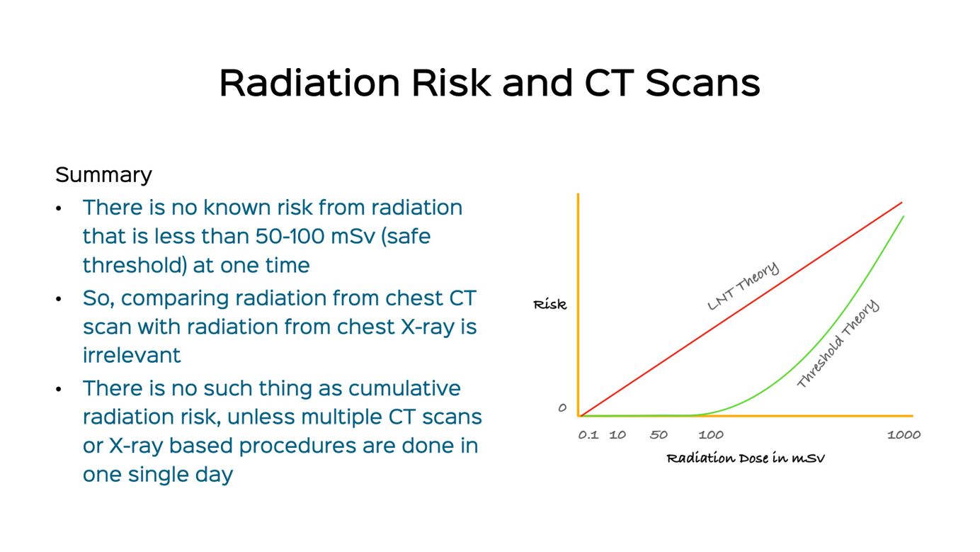
Snippet 03: Radiation Risk and CT Scan of the Chest Paid Members Public
126 years after the discovery of X-rays, there is no definitive data that radiation from CT scans increases the future risk of cancer.

Case 13: Hyperlucent Hemithorax in a 3-Years Old Paid Members Public
Hyperlucent hemithorax in a 3-years old

Snippet 02: Centrilobular / Bronchocentric Nodules Paid Members Public
A short discussion on centrilobular / bronchocentric nodules - how to diagnosis and characterize.
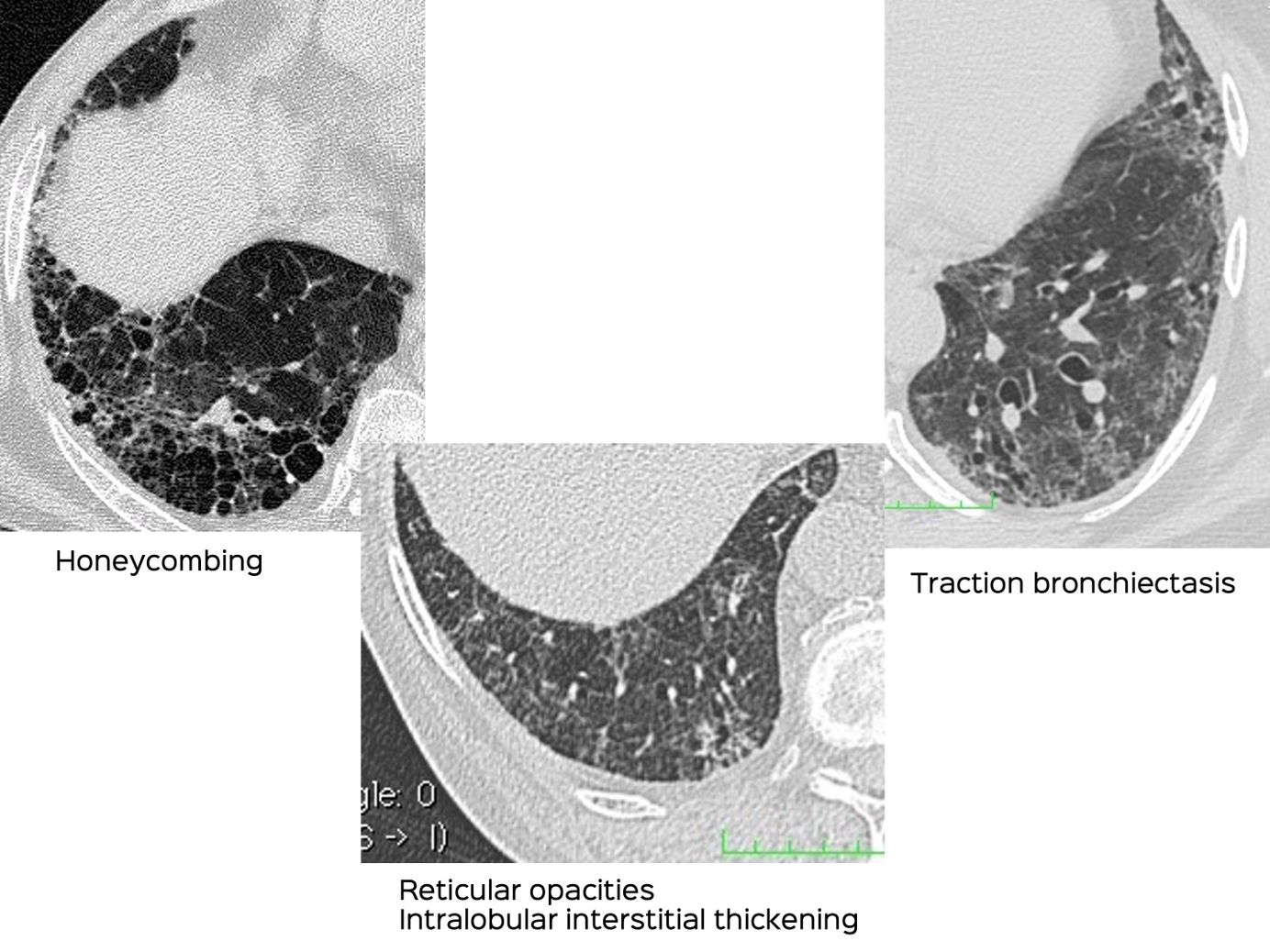
Snippet 01: Differentiating Traction Bronchiectasis from Honeycombing Paid Members Public
Many radiologists & physicians have difficulty differentiating traction bronchiectasis from honeycombing. The post and video explain how to do this.

CT Chest for Covid-19 - Indications - Position Statement / White Paper from Society of Chest Imaging & Intervention - SCII Paid Members Public
Position / white paper from the Society of Chest Imaging and Intervention (SCII) on the use of CT scan in Covid-19

Case 12: Tropical Pulmonary Eosinophilia Paid Members Public
How to suspect tropical pulmonary eosinophilia in acute/subacute settings with diffuse lung disease, especially ill-defined bronchocentric nodules.

Case 11: The Tragi-Comic Consequences of a Poor Quality Scan Paid Members Public
62-years old man diagnosed to have Covid-19 on an expiratory scan CT scan and the consequences thereafter. It is necessary to have a supine inspiratory scan at the least in all patients if expiratory and prone are not possible.

Case 10: Non-Resolving Progressive Opacities, Organizing Pneumonia and a Diagnosis After 3 Years Paid Members Public
67-years old lady who presented in 2017 with an organizing pneumonia (OP) pattern, and then 3 years later with significant progression
