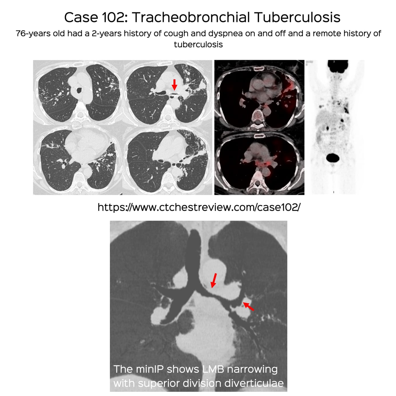
Case 102: Tracheobronchial Tuberculosis

It is important to recognize tracheobronchial TB on CT scans

Bhavin Jankharia

New One-Time, Lifetime Subscription

Please check out this page to see the changes in subscription models.
Payment
To make the site more accessible, we have decided to remove the yearly subscription and keep only a one-time, lifetime payment to get access to all content at www.ctchestreview.com and www.ctbiopsy.com. These sites are linked and hence one payment gives access to both sites, but the

Current Post

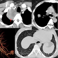
This 76-years old had a 2-years history of cough and dyspnea on and off and a remote history of tuberculosis.

A CT scan showed left main bronchus narrowing, for which a bronchoscopy was done and BAL showed likely malignant cells.

Repeat biopsy showed fibrosis with granulomatous disease but because of the activity, despite a negative microbiology she was put her on anti-TB treatment.

The short video below discusses the case, and then tracheobronchial tuberculosis, with four other cases.

Discussion Video


Table of Contents

Index and Table of Contents
Technical and Practice Issues * Case 11: 62-years old misdiagnosed to have interstitial lung disease - mid-inspiratory and expiratory scans * Snippet 03: Radiation risk and CT chest * Case 23: 60-years old - 40-pack years smoker - right upper lobe nodule - resolved using “mean” reconstructions…

My Linktree of All My Posts - Radiology and Non- Radiology

bjankharia | Instagram, Facebook | Linktree
Radiologist, Writer, Atmasvasth

Previous Post

Lecture: Risk Stratification of Lung Nodules
What to do when faced with a solitary lung nodule.

My New Book including the Kindle Edition

The Book - Atmasvasth - A Guide to Ageing Healthfully
A 15-point guide to living long, healthy

Other Sites and Cases

The Overweight Myth: Why Overnutrition Is Better Than Undernutrition Every Single Time!
Why it is better to overnourished due to food availability than undernourished due to food scarcity, every single time
Case 155: Early Darkness
All about microvascular obstruction (MVO)
Case 149: Upper Buccal Space Biopsy with a Paramaxillary Approach
Suprahyoid head neck biopsies are usually simple, if the anatomy is understood well. Buccal space lesion biopsies are even simpler.
TuberculosisAirway Disease

Comments


Featured Posts

Case 108: Approaching a Patient with PH - It Shouldn’t Be So Difficult!

Case 107: Why LLMs Will Render Many Radiologists and their Reports Irrelevant…

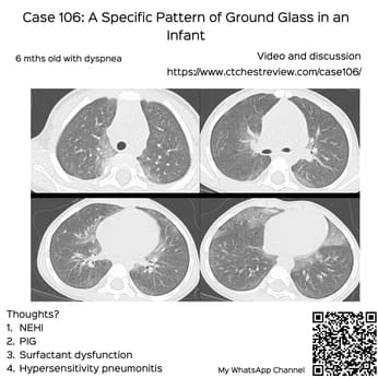
Case 106: A Specific Pattern of Ground Glass in an Infant

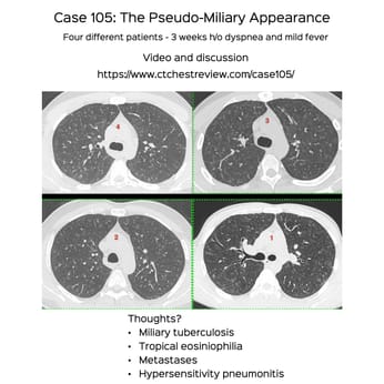
Case 105: The Pseudo-Miliary Appearance

Case 104: Popcorn is the Classic Appearance, but Not Always

Snippet 21: Approach to Right Medial Cardiophrenic Angle Lesions

Case 103: SFTP

Snippet 20: Practical Approach to Diffuse Cystic Lung Diseases

Case 102: Tracheobronchial Tuberculosis

Lecture: Risk Stratification of Lung Nodules

Case 101: When the Pattern of Lung Disease Gives a Clue to the Diagnosis

Snippet 19: The Various Uses of Prone Imaging

Case 100: CT of Covid-19...4 Years and 3 Months Later

Case 99: A Cleft Lung Puzzle

Lecture: Connective Tissue Disease ILDs

Case 98: Acute Increased Dyspnea in Idiopathic Pulmonary Fibrosis - First Considerations

Case 97: How to Identify and Diagnose Miliary TB as a Cause of ARDS

Case 96: When it Runs in the Family

Lecture: NSIP and OP

Lecture: How to Approach a Patient with Interstitial Lung Abnormality (ILA)

Case 95: When the Nodule is an Island

Lecture: Imaging of Sarcoidosis

Snippet 18: The Jellyfish and Multiple Pleural Tag Signs

Lecture: CT of Hypersensitivity Pneumonitis

Case 94: How Early is Early...

Case 93: The Asymmetric Chest Wall

Lecture: Lung Cancer Screening in a TB Endemic Country

REF-Cafe Roentgen Thoracic Thursday Lecture - XII - 26th Oct 2023: Mediastinum

Case 92: New Ground Glass in a Patient with a Fibrosing ILD

REF-Cafe Roentgen Thoracic Thursday Lecture - XI - 19th Oct 2023: Non-Resolving Opacity

Case 91: Bubble Lucencies, Central Lucencies, Bubbly Consolidation

Case 90: The Probable UIP Conundrum - Time Matters

REF-Cafe Roentgen Thoracic Thursday Lecture - X - 21st Sep 2023: Solitary Pulmonary Nodule - III - Subsolid Nodule

Case 89: Diaphragmatic Dysfunction – Eventration, Paralysis…Does it Matter

REF-Cafe Roentgen Thoracic Thursday Lecture - IX - 14th Sep 2023: Solitary Pulmonary Nodule - PFN, Juxtrapleural Nodule, Nodules in Smokers and Lung-RADs

REF-Cafe Roentgen Thoracic Thursday Lecture - VIII - 07th Sep 2023: Solitary Pulmonary Nodule > 8 mm

Snippet 17: How to Diagnose Subtle Progression of Interstitial Lung Disease at the Secondary Pulmonary Lobule Level

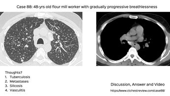
Case 88: Flour Mill Worker Disease

REF-Cafe Roentgen Thoracic Thursday Lecture - VII - 10th Aug 2023: Diffuse Cystic Lung Disease

REF-Cafe Roentgen Thoracic Thursday Lecture - VI - 03rd Aug 2023: Smoking Related Lung Diseases

Snippet 16: Inflammatory vs Fibrotic ILD

REF-Cafe Roentgen Thoracic Thursday Lecture - V - 20th July 2023: All About the Centrilobular Nodule

REF-Cafe Roentgen Thoracic Thursday Lecture - IV - 13th July 2023: Non-ILD Diffuse Lung Diseases

Case 87: Thymic Cyst or Cystic Thymoma

REF-Cafe Roentgen Thoracic Thursday Lecture - III - 29th June 2023: Non-Fibrosing ILDs

REF-Cafe Roentgen Thoracic Thursday Lecture - II - 22nd June 2023: Fibrosing ILDs - contd & Introduction to Non-Fibrosing ILDs

REF-Cafe Roentgen Thoracic Thursday Lecture - I: Fibrosing ILDs - 2023 06 15

Approach to Small Diffuse Lung Nodules - Lecture

Snippet 09: Variant Fibrosis

Snippet 08: Mosaic Attenuation

Snippet 04: Fibrotic NSIP vs Probable UIP

Snippet 02: Centrilobular / Bronchocentric Nodules

Snippet 01: Differentiating Traction Bronchiectasis from Honeycombing