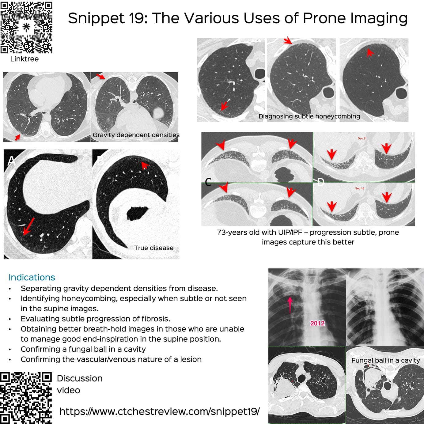
Snippet 19: The Various Uses of Prone Imaging

The many uses of prone imaging during CT chest examinations

Bhavin Jankharia

My Linktree of All My Posts - Radiology and Non- Radiology

bjankharia | Instagram, Facebook | Linktree
Radiologist, Writer, Atmasvasth

Current Post

This is a snippet discussing the various uses of prone imaging

Indications

  • Separating gravity dependent densities from disease.
  • Identifying honeycombing, especially when subtle or not seen in the supine images.
  • Evaluating subtle progression of fibrosis.
  • Obtaining better breath-hold images in those who are unable to manage good end-inspiration in the supine position.
  • Confirming a fungal ball in a cavity
  • Confirming the vascular/venous nature of a lesion

The short video below discusses these indications with examples

This post is for paying subscribers only

Subscribe

Already have an account? Log in


Featured Posts

Case 107: Why LLMs Will Render Many Radiologists and their Reports Irrelevant…

Case 106: A Specific Pattern of Ground Glass in an Infant

Case 105: The Pseudo-Miliary Appearance

Case 104: Popcorn is the Classic Appearance, but Not Always

Snippet 21: Approach to Right Medial Cardiophrenic Angle Lesions

Case 103: SFTP

Snippet 20: Practical Approach to Diffuse Cystic Lung Diseases

Case 102: Tracheobronchial Tuberculosis

Lecture: Risk Stratification of Lung Nodules

Case 101: When the Pattern of Lung Disease Gives a Clue to the Diagnosis

Snippet 19: The Various Uses of Prone Imaging

Case 100: CT of Covid-19...4 Years and 3 Months Later

Case 99: A Cleft Lung Puzzle

Lecture: Connective Tissue Disease ILDs

Case 98: Acute Increased Dyspnea in Idiopathic Pulmonary Fibrosis - First Considerations

Case 97: How to Identify and Diagnose Miliary TB as a Cause of ARDS

Case 96: When it Runs in the Family

Lecture: NSIP and OP

Lecture: How to Approach a Patient with Interstitial Lung Abnormality (ILA)

Case 95: When the Nodule is an Island

Lecture: Imaging of Sarcoidosis

Snippet: The Jellyfish and Multiple Pleural Tag Signs

Lecture: CT of Hypersensitivity Pneumonitis

Case 94: How Early is Early...

Case 93: The Asymmetric Chest Wall

Lecture: Lung Cancer Screening in a TB Endemic Country

REF-Cafe Roentgen Thoracic Thursday Lecture - XII - 26th Oct 2023: Mediastinum

Case 92: New Ground Glass in a Patient with a Fibrosing ILD

REF-Cafe Roentgen Thoracic Thursday Lecture - XI - 19th Oct 2023: Non-Resolving Opacity

Case 91: Bubble Lucencies, Central Lucencies, Bubbly Consolidation

Case 90: The Probable UIP Conundrum - Time Matters

REF-Cafe Roentgen Thoracic Thursday Lecture - X - 21st Sep 2023: Solitary Pulmonary Nodule - III - Subsolid Nodule

Case 89: Diaphragmatic Dysfunction – Eventration, Paralysis…Does it Matter

REF-Cafe Roentgen Thoracic Thursday Lecture - IX - 14th Sep 2023: Solitary Pulmonary Nodule - PFN, Juxtrapleural Nodule, Nodules in Smokers and Lung-RADs

REF-Cafe Roentgen Thoracic Thursday Lecture - VIII - 07th Sep 2023: Solitary Pulmonary Nodule > 8 mm

Snippet 17: How to Diagnose Subtle Progression of Interstitial Lung Disease at the Secondary Pulmonary Lobule Level

Case 88: Flour Mill Worker Disease

REF-Cafe Roentgen Thoracic Thursday Lecture - VII - 10th Aug 2023: Diffuse Cystic Lung Disease

REF-Cafe Roentgen Thoracic Thursday Lecture - VI - 03rd Aug 2023: Smoking Related Lung Diseases

Snippet 16: Inflammatory vs Fibrotic ILD

REF-Cafe Roentgen Thoracic Thursday Lecture - V - 20th July 2023: All About the Centrilobular Nodule

REF-Cafe Roentgen Thoracic Thursday Lecture - IV - 13th July 2023: Non-ILD Diffuse Lung Diseases

Case 87: Thymic Cyst or Cystic Thymoma

REF-Cafe Roentgen Thoracic Thursday Lecture - III - 29th June 2023: Non-Fibrosing ILDs

REF-Cafe Roentgen Thoracic Thursday Lecture - II - 22nd June 2023: Fibrosing ILDs - contd & Introduction to Non-Fibrosing ILDs

REF-Cafe Roentgen Thoracic Thursday Lecture - I: Fibrosing ILDs - 2023 06 15

Approach to Small Diffuse Lung Nodules - Lecture

Snippet 09: Variant Fibrosis

Snippet 08: Mosaic Attenuation

Snippet 04: Fibrotic NSIP vs Probable UIP

Snippet 02: Centrilobular / Bronchocentric Nodules

Snippet 01: Differentiating Traction Bronchiectasis from Honeycombing