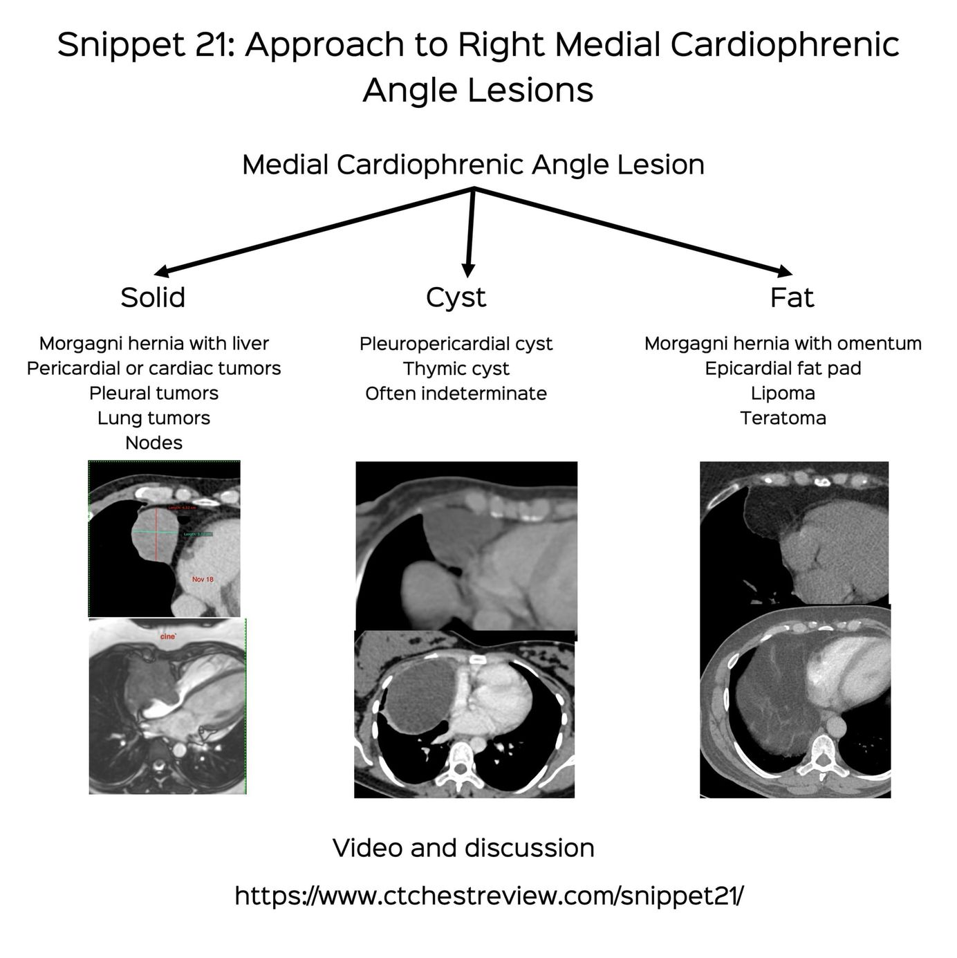
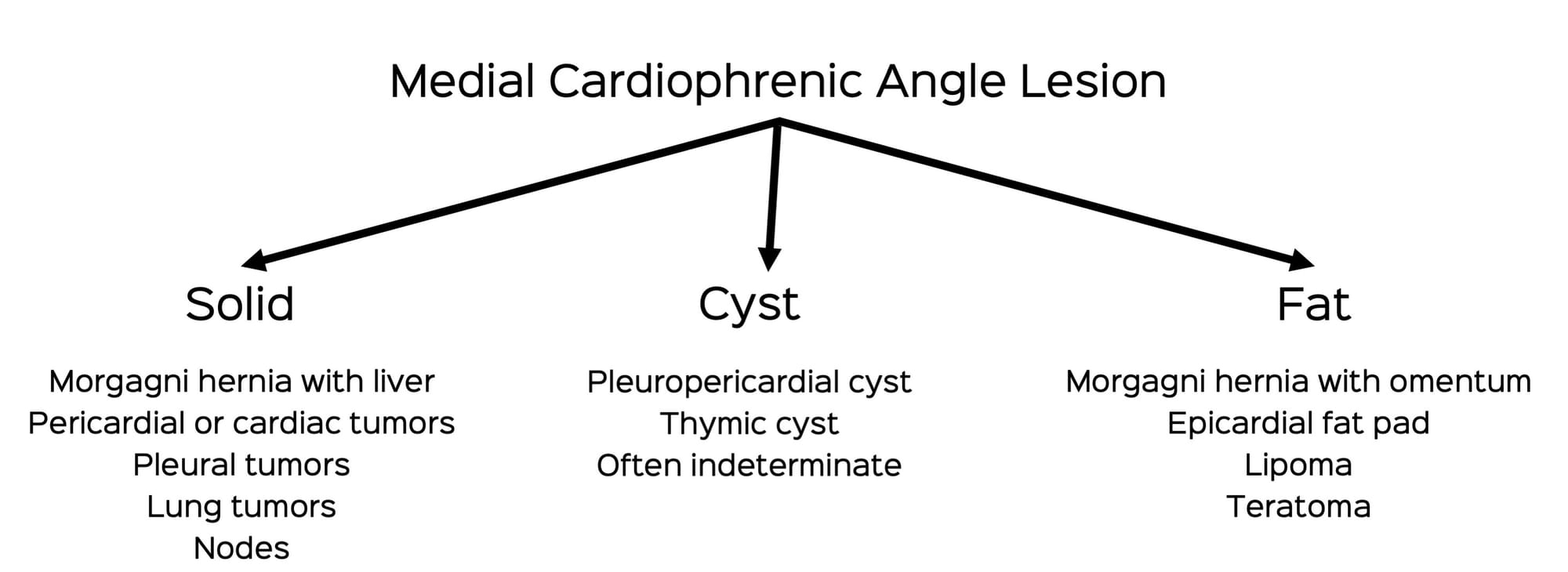
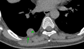
Snippet 21: Approach to Right Medial Cardiophrenic Angle Lesions
Right medial cardiophrenic angle lesions are a group of lesions that can be differentiate from one another based on their internal characteristics and location
New One-Time, Lifetime Subscription
Please check out this page to see the changes in subscription models.
Current Post
After the last post on SFTP, I realized I hadn't actually addressed the issue of right medial cardiophrenic angle lesions specifically, though there was one prior post on Morgagni hernias and a lecture on mediastinal lesions.
RMCPA (right medial cardiophrenic angle) lesions can be sorted out the same way as all mediastinal lesions, by first differentiating them into fat-containing, cystic or solid and then figuring out the origin (lung, pleura, mediastinum, heart).
This algorithm helps.
The video runs through the algorithm with examples and multiple cases illustrating how to to go about assessing RMCPA lesions.
This post is for paying subscribers only
SubscribeAlready have an account? Log in